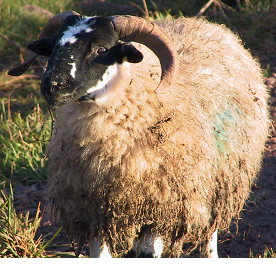

The eggs are then ingested by an intermediate host, where the eggs hatch in intestines and release oncospheres. brauni are shed in the feces of infected hosts into the environment. Ĭoenurosis in humans is rare and was not diagnosed until the twentieth century, with the first recorded cases by each Taenia species being: T. It was shown that the parasite could be transferred across species to and from dogs by Karl Theodor Ernst von Siebold and Friedrich Küchenmeister in the 1850s and the species was identified as Taenia multiceps (then called Coenurus cerebralis) in 1890. The cause of these cysts was identified as an animal parasite in 1780 by Nathanael Gottfried Leske and Johann August Ephraim Goeze. However, it was only in the 1600s that clearer behavioural and necropsy descriptions were recorded, including the chacteristic brain cysts and early surgical methods of removal. The texts of Hippocrates describe a nervous disease of sheep consistent with the symptoms of gid, comparing its symptoms to epilepsy and describing the accumulation of bad-smelling fluid in the brain. Humans cannot be definitive hosts for these species of tapeworms. The disease occurs mainly in sheep and other ungulates, but it can also occur in humans by accidental ingestion of tapeworm eggs.Īdult worms of these species develop in the small intestine of the definitive hosts (dogs, foxes and other canids), causing a disease from the group of taeniasis. It is caused by the coenurus, the larval stage of these tapeworms. Unless treated surgically, the animal will die ( Scott, 2007).Parasitic disease Different forms of coenurus in sheep and rabbits and an adult wormĬoenurosis, also known as caenurosis, coenuriasis, gid or sturdy, is a parasitic infection that develops in the intermediate hosts of some tapeworm species ( Taenia multiceps, T. As the cyst grows, the clinical signs progress to depression, unilateral blindness, circling, altered head position, in-coordination, paralysis ( Bussell et al., 1997) and recumbency. The earliest signs are often behavioral, with the affected animal tending to stand apart from the flock and react slowly to external stimuli. The time taken for the larvae to hatch, migrate and grow large enough to present nervous dysfunction varies from 2 to 6 months. Acute disease is an important differential diagnosis for Cerebrocortical necrosis (CCN).Ĭhronic coenurosis typically occurs in sheep of 16-18 months of age. Occasionally the signs are more severe and the animal may develop encephalitis, convulse and die within 4 – 5 days ( Scott, 2007). There is transient pyrexia, and relatively mild neurological signs such as listlessness and a slight head aversion. The signs are associated with an inflammatory and allergic reaction. Young lambs aged 6-8 weeks are most likely to show signs of acute disease. Acute coenurosis occurs during the migratory phase of the disease, usually about 10 days after the ingestion of large numbers of tapeworm eggs. The clinical signs of the coenurosis develop when the central nervous system (CNS) of the sheep is invaded by the cystic larval stage, or metacestode of the tapeworm.Ĭoenurosis can occur in both an acute and a chronic disease form. Usually the Coenurosis cerebralis cyst persists for the life of the intermediate host. The scolex (head of the tapeworm) embeds itself into the wall of the small intestine where it begins to grow, and shed new eggs. The life cycle is complete when the canine eats the raw infected brain, spinal cord or offal contaminated by the fluid from the ruptured cyst.The Coenurosis cerebralis matures into a thin-walled fluid-filled cyst about 5cm in diameter.The onchosphere develops into a metacestode larval stage called Coenurosis cerebralis.In goats the cysts can form in subcutaneous and muscular sites as well as the brain and spinal cord


The intermediate host is infected through ingestion of T.multiceps infection on a farm is significant as it confirms an unbroken sheep and dog life cycle, which in turn implies the existence of more important tapeworms such as Echinococcus granulosus. This dog / sheep tapeworm usually infects sheep and forms cysts in the lungs and liver, which if consumed by humans will cause a very serious disease that is very difficult to treat. Canine hosts shed tapeworm eggs in their feces which contaminates the pasture for the intermediate host to ingest. Dogs and other canines such as foxes, coyotes and jackals are the definitive hosts of the tapeworm Taenia multiceps.


 0 kommentar(er)
